- The University
- Studying
-
Research
- Profile
- Infrastructure
- Cooperations
- Services
-
Career
- Med Uni Graz as an Employer
- Educational Opportunities
- Work Environment
- Job openings
-
Diagnostics
- Patients
- Referring physicians
-
Health Topics
- Health Infrastructure
Bioimaging & Flow Cytometry
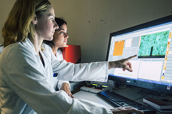
The Core Facilities Bioimaging and Flow Cytometry offers analyses of cells based on colorimetric, fluorescent, and luminescent detection by using microscopy, flow cytometry, and spectrophotometry techniques. These services enable for instance life-cell imaging/cell monitoring and high content screening of adherently growing cells. The established methods make it possible to identify around 200 different cell types in the human body with the help of specific “markers”. Isolated cell populations can be examined with regard to their function and reaction to changed environmental conditions or the mode of action of substances, drugs, etc.

Service
- (Confocal) light microscopy
- Measurement of cell membrane / tissue stiffness using atomic force microscopy
- Long-term cell observation
- Cell characterization using flow cytometry and cell sorting
- Cell physiological function tests (cytotoxicity, genotoxicity, immunotoxicity, etc.)
Infrastructure
The core facilities are equipped with the following devices and technologies:
- Confocal Laser Scan Microscopes (LSM510 Meta, Nikon A1R)
- High Content Screening (HCS Nikon)
- Atomic Force Microscopy (Flex-BIO, FLEX-ANA)
- Multicolor Flow Cytometry (CytoFLEX S, CytoFLEX LX)
- Cell Sorting (FACSARia IIIu)
- BioPlex-200, NanoSight NS300
Contact Bioimaging
Contact Bioimaging
Scientific Director
Assoz. Prof. Priv.-Doz Mag.Dr. Roland Malli
Assoz. Prof. Priv.-Doz Mag.Dr. Roland Malli
T: +43 316 385 71956


